This is an open source design for a smartphone camera microscope which can be customized, downloaded and 3D printed.
A team of researchers at RMIT University in Australia have developed a 3D printable clip-on microscope for smartphones. The design of the microscope has been shared on the Centre for Nanoscale BioPhotonics website.
Led by Anthony Orth, a research officer at the centre at RMIT University, the device has been developed to allow anyone, from students to medical staff to people at home, to take a closer look at things invisible to the naked eye.
The solution also means that the microscope can be used in situations where laboratory equipment may not be available. This could be hugely beneficial for application in less developed countries to help detect malaria or other blood borne parasites.
Orth explains in a post for The Conversation:
“What we’re hoping is that our design, or something like it, gets used for ultra simple, cheap and robust mobile phone based devices – be it for medical diagnostics in underserved areas such as the remote Australian outback and central Africa, or monitoring microorganism populations in local water sources.”
The researchers are also hoping that the final design can be optimized further to suit different people’s needs.
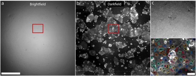
Bright and dark-field images taken with clip-on microscope. (Image: Nature)
Bright and dark-field microscopy possibilities
Orth and his team have proven that a smartphone already offers all the necessary parts to make a usable microscope. All that is missing is the magnification, which can be simply stuck on.
Samples also require illumination, which was achieved by using the smartphone’s internal flash. The challenge was to point the flash in the right direction to be able to shine through a sample and then into a camera.
Traditionally, this requires the use of prisms or mirror, but Orth and his team were able to diffuse the smartphone’s flash off of regular plastic. The clip-on design includes an assortment of tunnels to confine light and point it to the camera.
In addition to using the camera’s flash for a bright-field microscopy experience, the researchers also demonstrated that the sunlight could be used for illumination for a process called dark-field microscopy.
Finally, Orth recommends using Formlabs range of stereographic 3D printers to fabricate the design in black resin.
Source: Nature
Zooplankton using bright-field microscopy. (Image: Nature)
Website: LINK


